Chapter 20 Locomotion and Movements
Class 11th Biology NCERT Book Solution
NCERT Solutions For Class 11 Biology Locomotion and Movement
NCERT TEXTBOOK QUESTIONS FROM SOLVED
1. Draw the diagram of a
sarcomere of skeletal muscle showing different regions.
Solution: 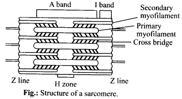
2. Define sliding filament
theory of muscle
contraction.
Solution: According to
sliding filament theory of muscle contraction, the actin and myosin filaments
slide past each other with the help of cross-bridges to reduce the length of the
sarcomeres.
3. Describe the important
steps in muscle
contraction.
Solution: Mechanism of
muscle contraction is explainei by sliding filament theory which states that
contraction of a muscle fibre takes place by the sliding of the thin filaments
over the th’ ck filaments. As a nerve impulse reaches the terminal end of the
axon, synaptic vesicles fuse with the axon membrane and release a chemical
transmitter, acetylcholine and binds to receptor sites of the motor end plate.
When depolarization of the motor end plate reaches a certain level, it creates
an action potential. An action potential (impulse) passes from the motor end
plate over the sarcolemma and then into the T-tubules and sarcoplasmic reticulum
and stimulates the sarcoplasmic reticulum to release calcium ions into the
sarcoplasm. The calcium ions bind to troponin causing a change in its shape and
position. This in turn alters shape and the position of tropomyosin, to which
troponin binds. This shift exposes the active sites on the F-actin molecules.
Myosin cross-bridges are then able to bind to these active sites. The heads of
myosin molecules project laterally from thick myofilaments towards the
surrounding thin myofilaments. These heads are called cross bridges. The head of
each myosin molecule contains an enzyme mysoin ATPase. In the presence of myosin
ATPase,Ca++ and Mg++ ions, ATP breaks down into ADP and
inorganic phosphate, releasing energy in the head.
Energy from ATP causes
energized myosin cross bridges to bind to actin.
The energized cross-bridges move, causing thin myofilaments
to slide along the thick myofilaments.
4. Write true or false. If
false change the statement so that it is true.
(a) Actin is present in thin filament.
(b) H-zone of striated muscle fibre represents both thick and thin
filaments.
(c) Human skeleton has 206 bones.
(d) There are 11 pairs of ribs in man.
(e) Sternum is present on the ventral side of the
body.
Solution: (a) True
(b) False –
H-Zone of striated muscle fibres represents only thick filaments.
(c)
True
(d) False – There are 12 pairs of ribs in man.
(e) True
5. Write the differences
between:
(a)
Actin and Myosin
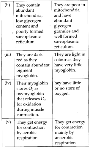
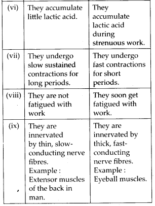
(b) Red and White
muscles
(c)
Pectoral and Pelvic
girdle
Solution: (a) Actin filaments
and myosin filaments can be differentiated as follows: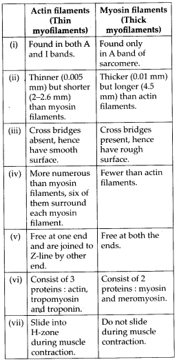
(b) Differences between red muscle fibres and white muscle
fibres are given in the following table:
(c) Differences between pectoral and pelvic girdles are given
in the following table: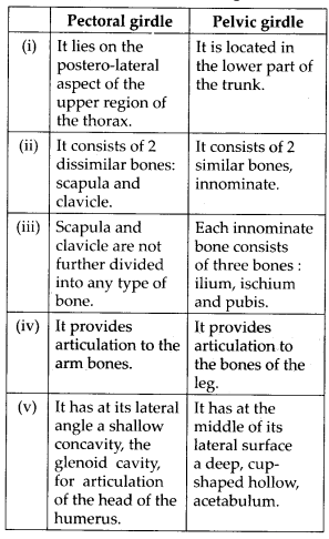
6. Match Column I with Column
II:
Column I
Column II
(a) Smooth muscle (i)
Myoglobin
(b) Tropomyosin (ii) Thin
filament
(c)
Red muscle (iii)
Sutures
(d)
Skull
(iv) Involuntary
Solution.(a)
– (iv), (b)-(ii), (c)-(i), (d)-(iii)
7. What are the different
types of movements exhibited by the cells of human
body?
Solution: The cells of human body
show three types of movements: amoeboid, ciliary and muscular.
Amoeboid
movements: These are found in leucocytes of blood and phagocytes of certain body
organs. In such cells, movements are brought with the help of temporary
finger-like cytoplasmic projections, called pseudopodia or false feet. So it is
also called pseudopodial movement. These pseudopodia are formed by flow of
cytoplasm, called cyclosis (simplest form of movement), and cytoskeletal
structures like microfilaments.
Ciliary movements: Large number of our
internal tubular organs are lined by ciliated epithelium. For instance, the
cilia of the cells lining the trachea, oviducts and vasa efferentia propel dust
particles, eggs and sperms respectively by their coordinated movements in
specific directions in these organs. Muscular movements: These are brought about
by the action of skeleton, joints and muscles. These are of two types: movements
of body parts and locomotion.
8. How do you distinguish
between a skeletal muscle and a cardiac
muscle?
Solution: We can distinguish
between a skeletal muscle and a cardiac muscle on the basis of the features
discussed in the following table: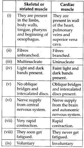
9. Name the type of joint
between the following:
(a) atlas/axis
(b) carpal/metacarpal of
thumb
(c)
between phalanges
(d)
femur/acetabulum
(e) between cranial
bones
(f)
between pubic bones in the pelvic
girdle
Solution: (a) Pivot joint
(b)
Saddle joint
(c) Hinge joint
(d) Ball and socket joint
(e) Fibrous
joint
(f) Cartilaginous joint
10. Fill in the blank
spaces:
(a)
All mammals (except a few) have……. cervical vertebra.
(b) The number of phalanges in each
limb of human is…….
(c) Thin filament of myofibril
contains two ‘F’ actins and two other proteins
namely…….and…….
(d) In a muscle fibre
Ca++ is stored in …….
(e)…….and…….pairs of ribs are called floating ribs.
(f) The human cranium is made of…….
bones.
Solution: (a) 7
(b) 14
(c)
tropomyosin, troponin
(d) sarcoplasmic reticulum
(e) 11th and 12th
(f)
8